

-
摘要: 深度学习(Deep learning,DL),特别是深度卷积神经网络(Convolutional neural networks,CNNs),能够从医学图像大数据中自动学习提取隐含的疾病诊断特征,近几年已迅速成为医学图像分析研究热点.本文首先简述医学图像分析特点;其次,论述深度学习基本原理,总结深度CNNs在医学图像分析中的分类、分割框架;然后,分别论述深度学习在医学图像分类、检测、分割等各应用领域的国内外研究现状;最后,探讨归纳医学图像分析深度学习方法挑战及其主要应对策略和开放的研究方向.Abstract: Deep learning (DL) algorithms, such as convolutional neural networks (CNNs), can automatically extract hidden disease diagnosis features from medical image data, and are being used to analyze medical images now. We review most of the deep learning methods for medical image analysis. Firstly, we introduce the characteristics of medical image analysis briefly. Then, we analyze the principles of deep learning, highlight the popular CNNs and summarize the frameworks of image classification and segmentation. Thirdly, we describe the state-of-the-art of the medical image analysis methods based on deep learning. Finally, we discuss the challenges and practicable strategies in deep learning for medical image analysis, as well as open research.1) 本文责任编委 桑农
-
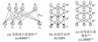
图 2 受限玻尔兹曼机RBM及基于RBM的深度网络
Fig. 2 Restricted Boltzmann machine (RBM) and deep networks based RBM
表 1 基于CNN的计算机视觉分类任务经典框架
Table 1 Classical CNN frameworks for computer vision classification tasks
网络结构 特点 备注 LeNet[9] 多个卷积层和子采样层 美国手写数字识别 AlexNet[60] 提出了ReLU和Dropout 刷新了2012年ImageNet ILSVRC物体分类竞赛的世界纪录 VGGNet[62] 提出采用小卷积核实现更深的网络以及多尺度融合 获ILSVRC 2014定位任务冠军、分类任务亚军 GoogleNet[65] 22层网络, 多个Inception结构串联 获ILSVRC 2014分类和检测任务冠军 ResNet[14] 提出了残差网络, 引入跳转连接, 深达152层 2015年ILSVRC物体检测与物体识别竞赛冠军 Inception ResNet[67] Inception结构与Residual Net结合 可获得与ResNet相当的性能, 但收敛速度加快 FCN[68] 密集性预测, 实现了像素级分类 避免了图像块之间的重叠而导致重复卷积计算的问题 DenseNet[70] 任何两层之间都有直接的连接 缓解梯度消失, 强化特征传播, 支持特征重用, 并降低网络参数数量 SqueezeNet[72] 简化网络结构和减少网络参数 仅需1/50的AlexNet参数量即可达到了AlexNet相同的精度 DCNN[73] 提出可变形深度卷积神经网络 增强了网络对于几何变换的建模能力 DPN[71] 结合了ResNet和DenseNet优势 基于DPN的团队取得2017年ILSVRC物体检测与物体识别竞赛冠军 SENet[74] 学习每个特征通道的重要程度, 强化有用特征 2017年ILSVRC图像分类任务竞赛冠军  下载: 导出CSV
表 2 脑瘤分割方法比较(使用BRATS数据集验证)
Table 2 Comparison of methods for brain tumor segmentation (validation on BRATS database)
作者 方法 DICE 总肿瘤区 核心肿瘤区 活性肿瘤区 专家评定 医学训练和经验 0.88 0.93 0.74 Urban[174] 多模态输入, 训练3D CNN 0.87 0.77 0.73 Zikic[175] 将3D立方体图像块转换成2D图像块, 训练2D CNN网络 0.837 0.736 0.69 Havaei[82] 2D多模态输入, 双路径级联CNN架构, 综合了局部细节和更全局信息 0.88 0.79 0.73 Pereira[176] 3×3的小的小卷积核, 更多的CNN层数和非线性运算, 更少的滤波器权重 0.88 0.83 0.77 Kamnitsas[168] 采用深度为11层的小滤波器3D CNN的双路径网络框架 0.898 0.75 0.721
下载: 导出CSV
-
[1] Bibault J E, Giraud P, Burgun A. Big data and machine learning in radiation oncology:state of the art and future prospects. Cancer Letters, 2016, 382(1):110-117 doi: 10.1016/j.canlet.2016.05.033 [2] Suzuki K, Zhou L P, Wang Q. Machine learning in medical imaging. Pattern Recognition, 2017, 63:465-467 doi: 10.1016/j.patcog.2016.10.020 [3] Hubel D H, Wiesel T N. Receptive fields of single neurones in the cat0s striate cortex. The Journal of Physiology, 1959, 148(3):574-591 doi: 10.1113/jphysiol.1959.sp006308 [4] Fukushima K, Miyake S, Ito T. Neocognitron:a neural network model for a mechanism of visual pattern recognition. IEEE Transactions on Systems, Man, and Cybernetics, 1983, SMC-13(5):826-834 doi: 10.1109/TSMC.1983.6313076 [5] Rumelhart D E, Hinton G E, Williams R J. Learning representations by back-propagating errors. Nature, 1986, 323(6088):533-536 doi: 10.1038/323533a0 [6] LeCun Y, Boser B, Denker J S, Henderson D, Howard R E, Hubbard W, Jackel L D. Backpropagation applied to handwritten zip code recognition. Neural Computation, 1989, 1(4):541-551 doi: 10.1162/neco.1989.1.4.541 [7] Hinton G E, Osindero S, Teh Y W. A fast learning algorithm for deep belief nets. Neural Computation, 2006, 18(7):1527-1554 doi: 10.1162/neco.2006.18.7.1527 [8] Hinton G E, Salakhutdinov R R. Reducing the dimensionality of data with neural networks. Science, 2006, 313(5786):504-507 doi: 10.1126/science.1127647 [9] Lecun Y, Bottou L, Bengio Y, Haffner P. Gradient-based learning applied to document recognition. Proceedings of the IEEE, 1998, 86(11):2278-2324 doi: 10.1109/5.726791 [10] Graves A, Schmidhuber J. Offline handwriting recognition with multidimensional recurrent neural networks. In: Proceedings of the 21st International Conference on Neural Information Processing Systems. British Columbia, Canada: Curran Associates Inc., 2012. 545-552 [11] Cireşan D C, Meier U, Gambardella L M, Schmidhuber J. Deep, Big, Simple neural nets for handwritten digit recognition. Neural Computation, 2010, 22(12):3207-3220 doi: 10.1162/NECO_a_00052 [12] Graves A, Jaitly N. Towards end-to-end speech recognition with recurrent neural networks. In: Proceedings of the 31st International Conference on Machine Learning. Beijing, China: PMLR, 2014. 1764-1772 [13] Hinton G, Deng L, Yu D, Dahl G E, Mohamed A R, Jaitly N, Senior A, Vanhoucke V, Nguyen P, Sainath T N, Kingsbury B. Deep neural networks for acoustic modeling in speech recognition:the shared views of four research groups. IEEE Signal Processing Magazine, 2012, 29(6):82-97 doi: 10.1109/MSP.2012.2205597 [14] He K M, Zhang X Y, Ren S Q, Sun J. Deep residual learning for image recognition. In: Proceedings of the ÿon Computer Vision and Pattern Recognition (CVPR). Las Vegas, NV, USA: IEEE, 2016. 770-778 [15] Sun Y, Wang X G, Tang X O. Deep learning face representation from predicting 10, 000 classes. In: Proceedings of the 2014 IEEE Conference on Computer Vision and Pattern Recognition. Columbus, OH, USA: IEEE, 2014. 1891-1898 [16] Mnih V, Kavukcuoglu K, Silver D, Rusu A A, Veness J, Bellemare M G, Graves A, Riedmiller M, Fidjeland A K, Ostrovski G, Petersen S, Beattie C, Sadik A, Antonoglou I, King H, Kumaran D, Wierstra D, Legg S, Hassabis D. Human-level control through deep reinforcement learning. Nature, 2015, 518(7540):529-533 doi: 10.1038/nature14236 [17] Helping clinicians get patients from test to treatment, faster[Online], available: http://www.deepmind.com/health, March 14, 2017. [18] 张蕾, 章毅.大数据分析的无限深度神经网络方法.计算机研究与发展, 2016, 53(1):68-79 doi: 10.7544/issn1000-1239.2016.20150663Zhang Lei, Zhang Yi. Big data analysis by infinite deep neural networks. Journal of Computer Research and Development, 2016, 53(1):68-79 doi: 10.7544/issn1000-1239.2016.20150663 [19] LeCun Y, Bengio Y, Hinton G. Deep learning. Nature, 2015, 521(7553):436-444 doi: 10.1038/nature14539 [20] Collobert R, Weston J. A unified architecture for natural language processing: deep neural networks with multitask learning. In: Proceedings of the 25th International Conference on Machine Learning (ICML). Helsinki, Finland: ACM, 2008. 160-167 [21] Taigman Y, Yang M, Ranzato M, Wolf L. Deepface: closing the gap to human-level performance in face verification. In: Proceedings of the 2014 IEEE Conference on Computer Vision and Pattern Recognition. Columbus, OH, USA: IEEE, 2014. 1701-1708 [22] Szegedy C, Toshev A, Erhan D. Deep neural networks for object detection. In: Proceedings of the 2013 Neural Information Processing Systems (NIPS). Lake Tahoe Nevada, USA: NIPS, 2013. 2553-2561 [23] Russakovsky O, Deng J, Su H, Krause J, Satheesh S, Ma S, Huang Z H, Karpathy A, Khosla A, Bernstein M, Berg A C, Fei-Fei L. ImageNet large scale visual recognition challenge. International Journal of Computer Vision, 2015, 115(3):211-252 doi: 10.1007/s11263-015-0816-y [24] VOC 2012[Online], available: https://coursys.sfu.ca/2016sp-cmpt-733-g2/pages/voc2012, March 14, 2017. [25] Mitos-atypi-14[Online], available: http://mitos-atypia-14.grand-challenge.org/, March 14, 2017. [26] Schmidhuber J. Deep Learning in neural networks:an overview. Neural Networks, 2015, 61:85-117 doi: 10.1016/j.neunet.2014.09.003 [27] Goodfellow I, Bengio Y, Courville A. Deep Learning. Massachusetts:MIT Press, 2016. [28] 段艳杰, 吕宜生, 张杰, 赵学亮, 王飞跃.深度学习在控制领域的研究现状与展望.自动化学报, 2016, 42(5):643-654 http://www.aas.net.cn/CN/abstract/abstract18852.shtmlDuan Yan-Jie, Lv Yi-Sheng, Zhang Jie, Zhao Xue-Liang, Wang Fei-Yue. Deep learning for control:the state of the art and prospects. Acta Automatica Sinica, 2016, 42(5):643-654 http://www.aas.net.cn/CN/abstract/abstract18852.shtml [29] 管皓, 薛向阳, 安志勇.深度学习在视频目标跟踪中的应用进展与展望.自动化学报, 2016, 42(6):834-847 http://www.aas.net.cn/CN/abstract/abstract18874.shtmlGuan Hao, Xue Xiang-Yang, An Zhi-Yong. Advances on application of deep learning for video object tracking. Acta Automatica Sinica, 2016, 42(6):834-847 http://www.aas.net.cn/CN/abstract/abstract18874.shtml [30] 罗建豪, 吴建鑫.基于深度卷积特征的细粒度图像分类研究综述.自动化学报, 2017, 43(8):1306-1318 http://www.aas.net.cn/CN/abstract/abstract19105.shtmlLuo Jian-Hao, Wu Jian-Xin. A Survey on fine-grained image categorization using deep convolutional features. Acta Automatica Sinica, 2017, 43(8):1306-1318 http://www.aas.net.cn/CN/abstract/abstract19105.shtml [31] Wells Ⅲ W M. Medical image analysis-past, present, and future. Medical Image Analysis, 2016, 33:4-6 doi: 10.1016/j.media.2016.06.013 [32] Madabhushi A, Lee G. Image analysis and machine learning in digital pathology:challenges and opportunities. Medical Image Analysis, 2016, 33:170-175 doi: 10.1016/j.media.2016.06.037 [33] Shen D G, Wu G R, Suk H I. Deep learning in medical image analysis. Annual Review of Biomedical Engineering, 2017, 19:221-248 doi: 10.1146/annurev-bioeng-071516-044442 [34] Greenspan H, van Ginneken B, Summers R M. Deep learning in medical imaging:overview and future promise of an exciting new technique. IEEE Transactions on Medical Imaging, 2016, 35(5):1153-1159 doi: 10.1109/TMI.2016.2553401 [35] Weese J, Lorenz C. Four challenges in medical image analysis from an industrial perspective. Medical Image Analysis, 2016, 33:44-49 doi: 10.1016/j.media.2016.06.023 [36] Rueckert D, Glocker B, Kainz B. Learning clinically useful information from images:past, present and future. Medical Image Analysis, 2016, 33:13-18 doi: 10.1016/j.media.2016.06.009 [37] Litjens G, Kooi T, Bejnordi B E, Setio A A A, Ciompi F, Ghafoorian M, van der Laak J A W M, van Ginneken B, Sánchez C. A survey on deep learning in medical image analysis. Medical Image Analysis, 2017, 42(9):60-88 http://arxiv.org/abs/1702.05747 [38] Spampinato C, Palazzo S, Giordano D, Aldinucci M, Leonardi R. Deep learning for automated skeletal bone age assessment in X-ray images. Medical Image Analysis, 2016, 36:41-51. https://www.sciencedirect.com/science/article/pii/S1361841516301840 [39] Li Y, Liang W, Zhang Y L, An H B, Tan J D. Automatic lumbar vertebrae detection based on feature fusion deep learning for partial occluded C-arm X-ray images. In: Proceedings of the 38th Annual International Conference of the Engineering in Medicine and Biology Society (EMBC). Orlando, FL, USA: IEEE, 2016. 647-650. [40] Nasr-Esfahani E, Samavi S, Karimi N, Soroushmehr S M R, Ward K, Jafari M H, Felfeliyan B, Nallamothu B, Najarian K. Vessel extraction in X-ray angiograms using deep learning. In: Proceedings of the 38th International Conference of the Engineering in Medicine and Biology Society (EMBC). Orlando, FL, USA: IEEE, 2016. 643-646. [41] Dobbs H J, Parker R P. The respective roles of the simulator and computed tomography in radiotherapy planning:a review. Clinical Radiology, 1984, 35(6):433-439 doi: 10.1016/S0009-9260(84)80035-5 [42] Niessen W J. MR brain image analysis in dementia:from quantitative imaging biomarkers to ageing brain models and imaging genetics. Medical Image Analysis, 2016, 33:107-113 doi: 10.1016/j.media.2016.06.029 [43] Powell C, Schmidt M, Borri M, Koh D M, Partridge M, Riddell A, Cookd G, Bhide S A, Nutting C M, Harrington K J, Newbold K L. Changes in functional imaging parameters following induction chemotherapy have important implications for individualised patient-based treatment regimens for advanced head and neck cancer. Radiotherapy and Oncology, 2013, 106(1):112-117 doi: 10.1016/j.radonc.2012.09.009 [44] Bagci U, Udupa J K, Mendhiratta N, Foster B, Xu Z Y, Yao J H, Chen X J, Mollura D J. Joint segmentation of anatomical and functional images:applications in quantification of lesions from PET, PET-CT, MRI-PET, and MRI-PET-CT images. Medical Image Analysis, 2013, 17(8):929-945 doi: 10.1016/j.media.2013.05.004 [45] Shi J, Zhou S C, Liu X, Zhang Q, Lu M H, Wang T F. Stacked deep polynomial network based representation learning for tumor classification with small ultrasound image dataset. Neurocomputing, 2016, 194:87-94 doi: 10.1016/j.neucom.2016.01.074 [46] Carneiro G, Nascimento J C, Freitas A. The segmentation of the left ventricle of the heart from ultrasound data using deep learning architectures and derivative-based search methods. IEEE Transactions on Image Processing, 2012, 21(3):968-982 doi: 10.1109/TIP.2011.2169273 [47] Gao X T, Lin S, Wong T Y. Automatic feature learning to grade nuclear cataracts based on deep learning. IEEE Transactions on Biomedical Engineering, 2015, 62(11):2693-2701 doi: 10.1109/TBME.2015.2444389 [48] Zilly J, Buhmann J M, Mahapatra D. Glaucoma detection using entropy sampling and ensemble learning for automatic optic cup and disc segmentation. Computerized Medical Imaging and Graphics, 2017, 55:28-41 doi: 10.1016/j.compmedimag.2016.07.012 [49] Yu L Q, Chen H, Dou Q, Qin J, Heng P A. Automated melanoma recognition in dermoscopy images via very deep residual networks. IEEE Transactions on Medical Imaging, 2017, 36(4):994-1004 doi: 10.1109/TMI.2016.2642839 [50] Dhungel N, Carneiro G, Bradley A P. A deep learning approach for the analysis of masses in mammograms with minimal user intervention. Medical Image Analysis, 2017, 37:114-128 doi: 10.1016/j.media.2017.01.009 [51] Bourlard H, Kamp Y. Auto-association by multilayer perceptrons and singular value decomposition. Biological Cybernetics, 1988, 59(4-5):291-294 doi: 10.1007/BF00332918 [52] Shin H C, Orton M R, Collins D J, Doran S J, Leach M O. Stacked autoencoders for unsupervised feature learning and multiple organ detection in a pilot study using 4D patient data. IEEE Transactions on Pattern Analysis and Machine Intelligence, 2013, 35(8):1930-1943 doi: 10.1109/TPAMI.2012.277 [53] Vincent P, Larochelle H, Lajoie I, Bengio Y, Manzagol P A. Stacked denoising autoencoders:learning useful representations in a deep network with a local denoising criterion. The Journal of Machine Learning Research, 2010, 11(12):3371-3408 [54] Smolensky P. Information Processing in Dynamical Systems:Foundations of Harmony Theory. Massachusetts:MIT Press, 1986. 194-281 [55] Salakhutdinov R, Hinton G. Deep Boltzmann machines. Journal of Machine Learning Research, 2009, 5(2):1967-2006 https://www.mendeley.com/research-papers/deep-boltzmann-machines/ [56] Carreira-Perpinan M A, Hinton G E. On contrastive divergence learning. In: Proceedings of the 2005 Artificial Intelligence and Statistics. Bridgetown, Barbados: AISTATS, 2005. [57] Larochelle H, Bengio Y, Louradour J, Lamblin P. Exploring strategies for training deep neural networks. The Journal of Machine Learning Research, 2009, 10:1-40 http://www.mendeley.com/catalog/exploring-strategies-training-deep-neural-networks/ [58] Bengio Y, Simard P, Frasconi P. Learning long-term dependencies with gradient descent is difficult. IEEE Transactions on Neural Networks, 1994, 5(2):157-166 http://www.ncbi.nlm.nih.gov/pubmed/18267787 [59] Hochreiter S, Schmidhuber J. Long short-term memory. Neural Computation, 1997, 9(8):1735-1780 doi: 10.1162/neco.1997.9.8.1735 [60] Krizhevsky A, Sutskever I, Hinton G E. ImageNet classification with deep convolutional neural networks. In: Proceedings of the 2012 International Conference on Neural Information Processing Systems. Lake Tahoe, Nevada, USA: Curran Associates Inc., 2012. 1097-1105 [61] Farabet C, Couprie C, Najman L, LeCun Y. Learning hierarchical features for scene labeling. IEEE Transactions on Pattern Analysis and Machine Intelligence, 2013, 35(8):1915-1929 doi: 10.1109/TPAMI.2012.231 [62] Simonyan K, Zisserman A. Very deep convolutional networks for large-scale image recognition. arXiv: 1409. 1556, 2014. [63] He K M, Zhang X Y, Ren S Q, Sun J. Spatial pyramid pooling in deep convolutional networks for visual recognition. IEEE Transactions on Pattern Analysis and Machine Intelligence, 2015, 37(9):1904-1916 doi: 10.1109/TPAMI.2015.2389824 [64] Lin M, Chen Q, Yan S C. Network in network. arXiv: 1312. 4400, 2014. [65] Szegedy C, Liu W, Jia Y Q, Sermanet P, Reed S, Anguelov D, Erhan D, Vanhoucke V, Rabinovich A. Going deeper with convolutions. In: Proceedings of the 2015 IEEE Conference on Computer Vision and Pattern Recognition. Boston, MA, USA: IEEE, 2015. 1-9 [66] Szegedy C, Vanhoucke V, Ioffe S, Shlens J, Wojna Z. Rethinking the Inception architecture for computer vision. In: Proceedings of the ÿon Computer Vision and Pattern Recognition. Las Vegas, NV, USA: IEEE, 2016. 2818-2826 [67] Szegedy C, Ioffe S, Vanhoucke V, Alemi A A. Inception-v4, Inception-ResNet and the impact of residual connections on learning. arXiv: 1602. 07261, 2016. [68] Long J, Shelhamer E, Darrell T. Fully convolutional networks for semantic segmentation. In: Proceedings of the 2015 IEEE Conference on Computer Vision and Pattern Recognition. Boston, Massachusetts, USA: IEEE, 2015. 3431-3440 [69] Zagoruyko S, Komodakis N. Wide residual networks. In: Proceedings of the 2016 British Machine Vision Conference. York, UK: BMVC, 2016. 87. 1-87. 12 [70] Huang G, Liu Z, van der Maaten L, Weinberger K Q. Densely connected convolutional networks. In: Proceedings of the 2017 IEEE Conference on Computer Vision and Pattern Recognition. Hawaii, USA: IEEE, 2017. 2261-2269 [71] Chen Y P, Li J A, Xiao H X, Jin X J, Yan S C, Feng J S. Dual path network. arXiv: 1707. 01629, 2017. [72] Iandola F N, Han S, Moskewicz M W, Ashraf K, Dally W J, Keutzer K. SqueezeNet: AlexNet-level accuracy with 50x fewer parameters and < 0. 5 MB model size. arXiv: 1602. 07360, 2016. [73] Dai J F, Qi H Z, Xiong Y W, Li Y, Zhang G D, Hu H, Wei Y C. Deformable convolutional networks. arXiv: 1703. 06211, 2017. [74] Hu J, Shen L, Sun G. Squeeze-and-excitation networks. arXiv: 1709. 01507, 2017. [75] ILSVRC2016[Online], available: http://image-net.org/challenges/LSVRC/2016/results, October 30, 2017. [76] LSVRC2017[Online], available: http://image-net.org/challenges/LSVRC/2017/results, October 30, 2017. [77] Dai J F, Li Y, He K M, Sun J. R-FCN: object detection via region-based fully convolutional networks. arXiv: 1605. 06409. 2016. [78] Nexar challenge Ⅱ[Online], available: https://www.getnexar.com/challenge-2/, October 30, 2017. [79] Cireşan D C, Giusti A, Gambardella L M, Schmidhuber J. Deep neural networks segment neuronal membranes in electron microscopy images. In: Proceedings of the 25th International Conference on Neural Information Processing Systems. Lake Tahoe, Nevada: Curran Associates Inc., 2012. 2843-2851 [80] Sermanet P, Eigen D, Zhang X, Mathieu M, Fergus R, Lecun Y. Overfeat: integrated recognition, localization and detection using convolutional networks. arXiv: 1312. 6229, 2014. [81] Cireşan D C, Giusti A, Gambardella L M, Schmidhuber J. Mitosis detection in breast cancer histology images with deep neural networks. Medical Image Computing and Computer-Assisted Intervention. Nagoya, Japan: Springer, 2013. 411-418 [82] Havaei M, Davy A, Warde-Farley D, Biard A, Courville A, Bengio Y, Pal C, Jodoin P M, Larochelle H. Brain tumor segmentation with deep neural networks. Medical Image Analysis, 2017, 35:18-31 doi: 10.1016/j.media.2016.05.004 [83] Choi H, Jin K H. Fast and robust segmentation of the striatum using deep convolutional neural networks. Journal of Neuroscience Methods, 2016, 274:146-153 doi: 10.1016/j.jneumeth.2016.10.007 [84] Song Y Y, Zhang L, Chen S P, Ni D, Lei B Y, Wang T F. Accurate segmentation of cervical cytoplasm and nuclei based on multiscale convolutional network and graph partitioning. IEEE Transactions on Biomedical Engineering, 2015, 62(10):2421-2433 doi: 10.1109/TBME.2015.2430895 [85] Yang W, Chen Y Y, Liu Y B, Zhong L M, Qin G G, Lu Z T, Feng Q J, Chen W F. Cascade of multi-scale convolutional neural networks for bone suppression of chest radiographs in gradient domain. Medical Image Analysis, 2017, 35, 421-433 doi: 10.1016/j.media.2016.08.004 [86] Ronneberger O, Fischer P, Brox T. U-net: convolutional networks for biomedical image segmentation. Medical Image Computing and Computer-Assisted Intervention. Munich, Germany: Springer, 2015, 9351: 234-241 [87] Çiçek Ö, Abdulkadir A, Lienkamp S S, Brox T, Ron-neberger O. 3D U-Net: learning dense volumetric segmentation from sparse annotation. Medical Image Computing and Computer-Assisted Intervention. Athens, Greece: Springer, 2016, 9901: 424-432 [88] Dou Q, Yu L Q, Chen H, Jin Y M, Yang X, Qin J, Heng P A. 3D deeply supervised network for automated segmentation of volumetric medical images. Medical Image Analysis, 2017, 41:40-45 doi: 10.1016/j.media.2017.05.001 [89] Milletari F, Navab N, Ahmadi S A. V-Net: fully convolutional neural networks for volumetric medical image segmentation. In: Proceedings of the 4th International Conference on 3D Vision. Stanford, CA, USA: IEEE, 2016. 565-571 [90] Suk H I, Lee S W, Shen D G. The Alzheimer0s Disease Neuroimaging Initiative. Hierarchical feature representation and multimodal fusion with deep learning for AD/MCI diagnosis. NeuroImage, 2014, 101:569-582 doi: 10.1016/j.neuroimage.2014.06.077 [91] Suk H I, Lee S W, Shen D G, The Alzheimer0s Disease Neuroimaging Initiative. Latent feature representation with stacked auto-encoder for AD/MCI diagnosis. Brain Structure and Function, 2015, 220(2):841-859 doi: 10.1007/s00429-013-0687-3 [92] Liu S Q, Liu S D, Cai W D, Che H Y, Pujol S, Kikinis R, Feng D G, Fulham M J. Multimodal neuroimaging feature learning for multiclass diagnosis of Alzheimer0s disease. IEEE Transactions on Biomedical Engineering, 2015, 62(4):1132-1140 doi: 10.1109/TBME.2014.2372011 [93] ANDI. Sharing Alzheimer0s research data with the world[Online], available: http://adni.loni.usc.edu/, March 14, 2017. [94] Al Rahhal M M, Bazi Y, AlHichri H, Alajlan N, Melgani F, Yager R R. Deep learning approach for active classification of electrocardiogram signals. Information Sciences, 2016, 345:340-354 doi: 10.1016/j.ins.2016.01.082 [95] Abdel-Zaher A M, Eldeib A M. Breast cancer classification using deep belief networks. Expert Systems with Applications, 2016, 46:139-144 doi: 10.1016/j.eswa.2015.10.015 [96] Arevalo J, González F A, Ramos-Pollán R, Oliveira J L, Lopez M A G. Representation learning for mammography mass lesion classification with convolutional neural networks. Computer Methods and Programs in Biomedicine, 2016, 127:248-257 doi: 10.1016/j.cmpb.2015.12.014 [97] Kooi T, Litjens G, van Ginneken B, Gubern-Mérida A, Sánchez C I, Mann R, den Heeten A, Karssemeijer N. Large scale deep learning for computer aided detection of mammographic lesions. Medical Image Analysis, 2017, 35:303-312 doi: 10.1016/j.media.2016.07.007 [98] Xu Y, Mo T, Feng Q W, Zhong P L, Lai M D, Chang E I C. Deep learning of feature representation with multiple instance learning for medical image analysis. In: Proceedings of the 2014 IEEE International Conference on Acoustics, Speech and Signal Processing. Florence, Italy: IEEE, 2014. 1626-1630 [99] Gao X W, Hui R, Tian Z M. Classification of CT brain images based on deep learning networks. Computer Methods and Programs in Biomedicine, 2017, 138:49-56 doi: 10.1016/j.cmpb.2016.10.007 [100] Payan A, Montana G. Predicting Alzheimer0s disease: a neuroimaging study with 3D convolutional neural networks. arXiv: 1502. 02506, 2015. [101] Hosseini-Asl E, Gimel0farb G, El-Baz A. Alzheimer0s disease diagnostics by a deeply supervised adaptable 3D convolutional network. arXiv: 1607. 00556, 2016. [102] Abdi A H, Luong C, Tsang T, Allan G, Nouranian S, Jue J, Hawley D, Fleming S, Gin K, Swift J, Rohling R, Abolmaesumi P. Automatic quality assessment of echocardiograms using convolutional neural networks:feasibility on the apical four-chamber view. IEEE Transactions on Medical Imaging, 2017, 36(6):1221-1230 doi: 10.1109/TMI.2017.2690836 [103] Gao X H, Li W, Loomes M, Wang L Y. A fused deep learning architecture for viewpoint classification of echocardiography. Information Fusion, 2017, 36:103-113 doi: 10.1016/j.inffus.2016.11.007 [104] Anthimopoulos M, Christodoulidis S, Ebner L, Christe A, Mougiakakou S. Lung pattern classification for interstitial lung diseases using a deep convolutional neural network. IEEE Transactions on Medical Imaging, 2016, 35(5):1207-1216 doi: 10.1109/TMI.2016.2535865 [105] Kawahara J, Hamarneh G. Multi-resolution-tract CNN with hybrid pretrained and skin-lesion trained layers. International Workshop on Machine Learning in Medical Imaging. Athens, Greece: Springer, 2016. 164-171 [106] Jiao Z C, Gao X B, Wang Y, Li J. A deep feature based framework for breast masses classification. Neurocomputing, 2016, 197:221-231 doi: 10.1016/j.neucom.2016.02.060 [107] Tajbakhsh N, Suzuki K. Comparing two classes of end-toend machine-learning models in lung nodule detection and classification:MTANNs vs. CNNs. Pattern Recognition, 2016, 63:476-486 https://www.sciencedirect.com/science/article/pii/S0031320316302795 [108] Kallenberg M, Petersen K, Nielsen M, Ng A Y, Diao P F, Igel C, VachonC M, Holland K, Winkel R R, Karssemeijer N, Lillholm M. Unsupervised deep learning applied to breast density segmentation and mammographic risk scoring. IEEE Transactions on Medical Imaging, 2016, 35(5):1322-1331 doi: 10.1109/TMI.2016.2532122 [109] van Tulder G, de Bruijne M. Combining generative and discriminative representation learning for lung CT analysis with convolutional restricted Boltzmann machines. IEEE Transactions on Medical Imaging, 2016, 35(5):1262-1272 doi: 10.1109/TMI.2016.2526687 [110] Zhang Q, Xiao Y, Dai W, Suo J F, Wang C Z, Shi J, Zheng H R. Deep learning based classification of breast tumors with shear-wave elastography. Ultrasonics, 2016, 72:150-157 doi: 10.1016/j.ultras.2016.08.004 [111] Yang D, Zhang S T, Yan Z N, Tan C W, Li K, Metaxas D. Automated anatomical landmark detection ondistal femur surface using convolutional neural network. In: Proceedings of the 12th International Symposium on Biomedical Imaging. New York, NY, USA: IEEE, 2015. 17-21 [112] Chen H, Dou Q, Wang X, Qin J, Cheng J C Y, Heng P A. 3D fully convolutional networks for intervertebral disc localization and segmentation. Medical Imaging and Augmented Reality. Cham:Springer, 2016, 9805:375-382 doi: 10.1007/978-3-319-43775-0_34 [113] de Vos B D, Wolterink J M, de Jong P A, Viergever M A, Išgum I. 2D image classification for 3D anatomy localization: employing deep convolutional neural networks. In: Proceedings of the 9784, Medical Imaging 2016: Image Processing. San Diego, California, US: SPIE, 2016, 9784: Article No. 97841Y [114] Kong B, Zhan Y Q, Shin M, Denny T, Zhang S T. Recognizing end-diastole and end-systole frames via deep temporal regression network. In: Medical Image Computing and Computer-Assisted Intervention-MICCAI. Athens, Greece: Springer, 2016. 264-272 [115] Cai Y L, Landis M, Laidley D T, Kornecki A, Lum A, Li S. Multi-modal vertebrae recognition using Transformed Deep Convolution Network. Computerized Medical Imaging and Graphics, 2016, 51:11-19 doi: 10.1016/j.compmedimag.2016.02.002 [116] Lei J, Li G H, Tu D, Guo Q. Convolutional restricted Boltzmann machines learning for robust visual tracking. Neural Computing and Applications, 2014, 25(6):1383-1391 doi: 10.1007/s00521-014-1625-x [117] Lo S C B, Lou S L A, Lin J S, Freedman M T, Chien M V, Mun S K. Artificial convolution neural network techniques and applications for lung nodule detection. IEEE Transactions on Medical Imaging, 1995, 14(4):711-718 doi: 10.1109/42.476112 [118] Sirinukunwattana K, Raza S E A, Tsang Y W, Snead D R J, Cree I A, Rajpoot N M. Locality sensitive deep learning for detection and classification of nuclei in routine colon cancer histology images. IEEE Transactions on Medical Imaging, 2016, 35(5):1196-1206 doi: 10.1109/TMI.2016.2525803 [119] Li A N, Cheng J, Wong D W K, Liu J. Integrating holistic and local deep features for glaucoma classification. In: Proceedings of the 38th Annual International Conference of the Engineering in Medicine and Biology Society. Orlando, FL, USA: IEEE, 2016. 1328-1331 [120] Roth H R, Lu L, Seff A, Cherry K M, Hoffman J, Wang S J, Liu J M, Turkbey E, Summers R M. A new 2. 5D representation for lymph node detection using random sets of deep convolutional neural network observations. In: Proceedings of the 17th International Conference International Conference on Medical Image Computing and ComputerAssisted Intervention. Cham, Germany: Springer, 2013, 8673: 520-527 [121] Roth H R, Lu L, Liu J M, Yao J H, Seff A, Cherry K, Kim L, Summers R M. Improving computer-aided detection using convolutional neural networks and random view aggregation. IEEE Transactions on Medical Imaging, 2016, 35(5):1170-1181 doi: 10.1109/TMI.2015.2482920 [122] Roth H R, Yao J H, Lu L, Stieger J, Burns J E, Summers R M. Detection of sclerotic spine metastases via random aggregation of deep convolutional neural network classifications. Recent Advances in Computational Methods and Clinical Applications for Spine Imaging. Cham: Springer, 2015. 3-12 [123] Wang J, Ding H J, Bidgoli F A, Zhou B, Iribarren C, Molloi S, Baldi P. Detecting cardiovascular disease from mammograms with deep learning. IEEE Transactions on Medical Imaging, 2017, 36(5):1172-1181 doi: 10.1109/TMI.2017.2655486 [124] Quellec G, Charrière K, Boudi Y, Cochener B, Lamard M. Deep image mining for diabetic retinopathy screening. Medical Image Analysis, 2017, 39:178-193 doi: 10.1016/j.media.2017.04.012 [125] Teramoto A, Fujita H, Yamamuro O, Tamaki T. Automated detection of pulmonary nodules in PET/CT images:ensemble false-positive reduction using a convolutional neural network technique. Medical Physics, 2016, 43:2821-2827 doi: 10.1118/1.4948498 [126] Albarqouni S, Baur C, Achilles F, Belagiannis V, Demirci, Navab N. AggNet:deep learning from crowds for mitosis detection in breast cancer histology images. IEEE Transactions on Medical Imaging, 2016, 35(5):1313-1321 doi: 10.1109/TMI.2016.2528120 [127] Chen H, Yu L Q, Dou Q, Shi L, Mok V C T, Heng P A. Automatic detection of cerebral microbleeds via deep learning based 3D feature representation. In: Proceedings of the 12th International Symposium on Biomedical Imaging (ISBI). New York, NY, USA: IEEE, 2015. 764-767 [128] Dou Q, Chen H, Yu L Q, Zhao L, Qin J, Wang D F, Mok V C T, Shi L, Heng P A. Automatic detection of cerebral microbleeds from MR images via 3D convolutional neural networks. IEEE Transactions on Medical Imaging, 2016, 35(5):1182-1195 doi: 10.1109/TMI.2016.2528129 [129] Dou Q, Chen H, Yu L Q, Qin J, Heng P A. Multilevel contextual 3-D CNNs for false positive reduction in pulmonary nodule detection. IEEE Transactions on Biomedical Engineering, 2017, 64(7):1558-1567 doi: 10.1109/TBME.2016.2613502 [130] van Grinsven M J J P, van Ginneken B, Hoyng C B, Theelen T, Sanchez C I. Fast convolutional neural network training using selective data sampling:application to hemorrhage detection in color fundus images. IEEE Transactions on Medical Imaging, 2016, 35(5):1273-1284 doi: 10.1109/TMI.2016.2526689 [131] Xu J, Xiang L, Liu Q S, Gilmore H, Wu J Z, Tang J H, Madabhushi A. Stacked sparse autoencoder (SSAE) for nuclei detection on breast cancer histopathology images. IEEE Transactions on Medical Imaging, 2016, 35(1):119-130 doi: 10.1109/TMI.2015.2458702 [132] Masood A, Al-Jumaily A, Anam K. Self-supervised learning model for skin cancer diagnosis. In: Proceedings of the 7th International IEEE/EMBS Conference on Neural Engineering. Montpellier, USA: IEEE, 2015. 1012-1015 [133] Girshick R. Fast R-CNN. In: Proceedings of the 2015 IEEE International Conference on Computer Vision. Santiago, Chile: IEEE, 2015. 1440-1448 [134] Sarikaya D, Corso J J, Guru K A. Detection and localization of robotic tools in robot-assisted surgery videos using deep neural networks for region proposal and detection. IEEE Transactions on Medical Imaging, 2017, 36(7):1542-1549 doi: 10.1109/TMI.2017.2665671 [135] Twinanda A P, Shehata S, Mutter D, Marescaux J, Mathelin M D, Padoy N. EndoNet:a deep architecture for recognition tasks on laparoscopic videos. IEEE Transactions on Medical Imaging, 2016, 36(1):86-97 http://www.ncbi.nlm.nih.gov/pubmed/27455522 [136] Chen H, Wu L Y, Dou Q, Qin J, Li S, Cheng J Z, et al. Ultrasound standard plane detection using a composite neural network Framework. IEEE Transactions on Cybernetics, 2017, 47(6):1576-1586 doi: 10.1109/TCYB.2017.2685080 [137] Kumar N, Verma R, Sharma S, Bhargava S, Vahadane A, Sethi A. A dataset and a technique for generalized nuclear segmentation for computational pathology. IEEE Transactions on Medical Imaging, 2017, 36(7):1550-1560 doi: 10.1109/TMI.2017.2677499 [138] Xu Y, Jia Z P, Ai Y Q, Zhang F, Lai M D, Chang E I C. Deep convolutional activation features for large scale brain tumor histopathology image classification and segmentation. In: Proceedings of the 2015 IEEE International Conference on Acoustics, Speech and Signal Processing (ICASSP). South Brisbane, QLD, Australia: IEEE, 2015. 947-951 [139] Qaiser T, Sirinukunwattana K, Nakane K, Tsang Y W, Epstein D, Rajpoot N. Persistent homology for fast tumor segmentation in whole slide histology images. Procedia Computer Science, 2016, 90:119-124 doi: 10.1016/j.procs.2016.07.033 [140] Song Y Y, Tan E L, Jiang X D, Cheng J Z, Ni D, Chen S P, Lei B Y, Wang T F. Accurate cervical cell segmentation from overlapping clumps in pap smear images. IEEE Transactions on Medical Imaging, 2017, 36(1):288-300 doi: 10.1109/TMI.2016.2606380 [141] Fakhry A, Zeng T, Ji S W. Residual deconvolutional networks for brain electron microscopy image segmentation. IEEE Transactions on Medical Imaging, 2017, 36(2):447-456 doi: 10.1109/TMI.2016.2613019 [142] Chen H, Qi X J, Yu L Q, Heng P A. DCAN: deep contouraware networks for accurate gland segmentation. In: Proceedings of the ÿon Computer Vision and Pattern Recognition (CVPR). Las Vegas, NV, USA: IEEE, 2016. 2487-2496 [143] Xu Y, Li Y, Wang Y P, Liu M Y, Fan Y B, Lai M D, Chang E I C. Gland instance segmentation using deep multichannel neural networks. IEEE Transactions on Biomedical Engineering, 2017, 64(12):2901-2912 doi: 10.1109/TBME.2017.2686418 [144] Sirinukunwattana K, Pluim J P W, Chen H, Qi X J, Heng P A, Guo Y B, Wang L Y, Matuszewski B J, Bruni E, Sanchez U, Böhm A, Ronneberger O, Cheikh B B, Racoceanu D, Kainz P, Pfeiffer M, Urschler M, Snead D R J, Snead N M. Gland segmentation in colon histology images:the glas challenge contest. Medical Image Analysis, 2016, 35:489-502 http://www.ncbi.nlm.nih.gov/pubmed/27614792 [145] Xie Y P, Zhang Z Z, Sapkota M, Yang L. Spatial clockwork recurrent neural network for muscle perimysium segmentation. International Conference on Medical Image Computing and Computer-Assisted Intervention. Athens, Greece: Springer, 2016, 9901: 185-193 [146] Xing F Y, Xie Y P, Yang L. An automatic learning-based framework for robust nucleus segmentation. IEEE Transactions on Medical Imaging, 2016, 35(2):550-566 doi: 10.1109/TMI.2015.2481436 [147] Zhang W L, Li R J, Deng H T, Wang L, Lin W L, Ji S W, Shen D G. Deep convolutional neural networks for multi-modality isointense infant brain image segmentation. NeuroImage, 2015, 108:214-224 doi: 10.1016/j.neuroimage.2014.12.061 [148] Stollenga M F, Byeon W, Liwicki M, Schmidhuber J. Parallel multi-dimensional LSTM, with application to fast biomedical volumetric image segmentation. arXiv: 1506. 07452, 2015. [149] Cui Z P, Yang J, Qiao Y. Brain MRI segmentation with patch-based CNN approach. In: Proceedings of the 35th Chinese Control Conference. Chengdu, China: IEEE, 2016. 7026-7031 [150] Moeskops P, Viergever M A, Mendrik A M, de Vries L S, Benders M J N L, Išgum I. Automatic segmentation of MR brain images with a convolutional neural network. IEEE Transactions on Medical Imaging, 2016, 35(5):1252-1262 doi: 10.1109/TMI.2016.2548501 [151] Dolz J, Betrouni N, Quidet M, Kharroubi D, Leroy H A, Reyns N, Massoptier L, Vermandel M. Stacking denoising auto-encoders in a deep network to segment the brainstem on MRI in brain cancer patients:a clinical study. Computerized Medical Imaging and Graphics, 2016, 52:8-18 doi: 10.1016/j.compmedimag.2016.03.003 [152] Mansoor A, Cerrolaza J J, Idrees R, Biggs E, Alsharid M A, Avery R A, Linguraru M G. Deep learning guided partitioned shape model for anterior visual pathway segmentation. IEEE Transactions on Medical Imaging, 2016, 35(8):1856-1865 doi: 10.1109/TMI.2016.2535222 [153] Shakeri M, Tsogkas S, Ferrante E, Lippe S, Kadoury S, Paragios N, Kokkinos I. Sub-cortical brain structure segmentation using F-CNN0s. In: Proceedings of the 13th International Symposium on Biomedical Imaging (ISBI). Prague, Czech: IEEE, 2016. 269-272 [154] Moeskops P, Wolterink J M, van der Velden B H M, Gilhuijs K G A, Leiner T, Viergever M A, Išgum I. Deep learning for multi-task medical image segmentation in multiple modalities. Medical Image Computing and Computer-Assisted Intervention. Athens, Greece: Springer, 2016, 9901: 478-486 [155] Wu A, Xu Z Y, Gao M C, Buty M, Mollura D J. Deep vessel tracking: a generalized probabilistic approach via deep learning. In: Proceedings of the 13th International Symposium on Biomedical Imaging (ISBI). Prague, Czech: IEEE, 2016. 1363-1367 [156] Liskowski P, Krawiec K. Segmenting retinal blood vessels with deep neural networks. IEEE Transactions on Medical Imaging, 2016, 35(11):2369-2380 doi: 10.1109/TMI.2016.2546227 [157] Wang S L, Yin Y L, Cao G B, Wei B Z, Zheng Y J, Yang G P. Hierarchical retinal blood vessel segmentation based on feature and ensemble learning. Neurocomputing, 2015, 149:708-717 doi: 10.1016/j.neucom.2014.07.059 [158] Li Q L, Feng B W, Xie L P, Liang P, Zhang H S, Wang T F. A cross-modality learning approach for vessel segmentation in retinal images. IEEE Transactions on Medical Imaging, 2016, 35(1):109-118 doi: 10.1109/TMI.2015.2457891 [159] Avendi M R, Kheradvar A, Jafarkhani H. A combined deep-learning and deformable-model approach to fully automatic segmentation of the left ventricle in cardiac MRI. Medical Image Analysis, 2016, 30:108-119 doi: 10.1016/j.media.2016.01.005 [160] Ngo T A, Lu Z, Carneiro G. Combining deep learning and level set for the automated segmentation of the left ventricle of the heart from cardiac cine magnetic Resonance. Medical Image Analysis, 2017, 35:159-171 doi: 10.1016/j.media.2016.05.009 [161] Tan L K, Liew Y M, Lim E, McLaughlin R A. Convolutional neural network regression for short-axis left ventricle segmentation in cardiac cine MR sequences. Medical Image Analysis, 2017, 39:78-86 doi: 10.1016/j.media.2017.04.002 [162] Zhen X, Wang Z, Islam A, Bhaduri M, Chan I, Li S. Multi-scale deep networks and regression forests for direct bi-ventricular volume estimation. Medical Image Analysis, 2015, 30:120-129 http://www.sciencedirect.com/science/article/pii/S1361841515001024 [163] Roth H R, Farag A, Lu L, Turkbey E B, Ronald M. Deep convolutional networks for pancreas segmentation in CT imaging. In: Proceedings of 9413, Medical Imaging 2015: Image Processing. Orlando, Florida, USA: SPIE, 2015, 9413: Article No. 94131G [164] Roth H R, Lu L, Farag A, Shin H C, Liu J M, Turkbey E B, Summers R M. DeepOrgan: multi-level deep convolutional networks for automated pancreas segmentation. In: Proceedings of the 18th International Conference Medical Image Computing and Computer-Assisted Intervention-MICCAI 2015. Munich, Germany: Springer, 2015. 556-564 [165] Guo Y R, Gao Y Z, Shen D G. Deformable MR prostate segmentation via deep feature learning and sparse patch matching. IEEE Transactions on Medical Imaging, 2016, 35(4):1077-1089 doi: 10.1109/TMI.2015.2508280 [166] Korez R, Likar B, Pernuš F, Vrtovec T. Model-based segmentation of vertebral bodies from MR images with 3D CNNs. Medical Image Computing and Computer-Assisted Intervention. Athens, Greece: Springer, 2016. 433-441 [167] Poudel R P K, Lamata P, Montana G. Recurrent fully convolutional neural networks for multi-slice MRI cardiac segmentation. Reconstruction, Segmentation, and Analysis of Medical Images. Cham: Springer, 2016. 83-94 [168] Kamnitsas K, Ledig C, Newcombe V F J, Simpson J P, Kane A D, Menon D K, Rueckert D, Glocker B. Efficient multi-scale 3D CNN with fully connected CRF for accurate brain lesion segmentation. Medical Image Analysis, 2017, 36:61-78 doi: 10.1016/j.media.2016.10.004 [169] Chen L C, Papandreou G, Kokkinos I, Murphy K, Yuille. Semantic image segmentation with deep convolutional nets and fully connected CRFs. arXiv: 1412. 7062, 2014. [170] Gao M, Xu Z Y, Lu L, Wu A, Nogues I, Summers R M, Mollura D J. Segmentation label propagation using deep convolutional neural networks and dense conditional random field. In: Proceedings of the 13th International Symposium on Biomedical Imaging. Prague, Czech Republic: IEEE, 2016. 1265-1268 [171] Brosch T, Tang L Y W, Yoo Y, Li D K B, Traboulsee A, Tam R. Deep 3D convolutional encoder networks with shortcuts for multiscale feature integration applied to multiple sclerosis lesion segmentation. IEEE Transactions on Medical Imaging, 2016, 35(5):1229-1239 doi: 10.1109/TMI.2016.2528821 [172] BRATS2015[Online], available: https://www.smir.ch/BRATS/Start2015, March 14, 2017. [173] Wang C H, Yan X C, Smith M, Kochhar K, Rubin M, Warren S M, Wrobel J, Lee H. A unified framework for automatic wound segmentation and analysis with deep convolutional neural networks. In: Proceedings of the 37th Annual International Conference of the IEEE Engineering in Medicine and Biology Society. Milan, Italy: IEEE, 2015. 2415-2418 [174] Urban G, Bendszus M, Hamprecht F A, Kleesiek J. Multimodal brain tumor segmentation using deep convolutional neural networks. In: Proceedings of the 2014 MICCAI Workshop on Multimodal Brain Tumor Segmentation Challenge. Boston, Massachusetts, USA: Springer, 2014. 31-35 [175] Zikic D, Ioannou Y, Brown M, Criminisi A. Segmentation of brain tumor tissues with convolutional neural networks. In: Proceedings of the 2014 MICCAI Workshop on Multimodal Brain Tumor Segmentation Challenge. Boston, Massachusetts, USA: Springer, 2014. 36-39 [176] Pereira S, Pinto A, Alves V, Silva C A. Brain tumor segmentation using convolutional neural networks in MRI images. IEEE Transactions on Medical Imaging, 2016, 35(5):1240-1251 doi: 10.1109/TMI.2016.2538465 [177] Carneiro G, Nascimento J, Bradley A P. Unregistered multiview mammogram analysis with pre-trained deep learning models. Medical Image Computing and ComputerAssisted Intervention. Munich, Germany: Springer, 2015. 652-660 [178] Bar Y, Diamant I, Wolf L, Greenspan H. Deep learning with non-medical training used for chest pathology identification. In: Proceedings of 9414, Medical Imaging 2015: Computer-Aided Diagnosis. Orlando, Florida, USA: SPIE, 2015, 9414: Article No. 94140V [179] Bar Y, Diamant I, Wolf L, Lieberman S, Konen E, Greenspan H. Chest pathology detection using deep learning with non-medical training. In: Proceedings of the 12th International Symposium on Biomedical Imaging. New York, NY, USA: IEEE, 2015. 294-297 [180] Ginneken B V, Setio A A A, Jacobs C, Ciompi F. Off-theshelf convolutional neural network features for pulmonary nodule detection in computed tomography scans. In: Proceedings of the 12th International Symposium on Biomedical Imaging. New York, NY, USA: IEEE, 2015. 286-289 [181] Shin H C, Roth H R, Gao M C, Lu L, Xu Z Y, Nogues I, Yao J H, Mollura D, Summers R M. Deep convolutional neural networks for computer-aided detection:CNN architectures, dataset characteristics and transfer learning. IEEE Transactions on Medical Imaging, 2016, 35(5):1285-1298 doi: 10.1109/TMI.2016.2528162 [182] Chatfield K, Simonyan K, Vedaldi A, Zisserman A. Return of the devil in the details: delving deep into convolutional nets. In: Proceedings of the 2014 British Machine Vision Conference. Nottingham, UK: University of Nottingham, 2014. [183] Ciompi F, de Hoop B, van Riel S J, Chung K, Scholten E T, Oudkerk M, de Jong P A, Prokop M, van Ginneken B. Automatic classification of pulmonary peri-fissural nodules in computed tomography using an ensemble of 2D views and a convolutional neural network out-of-the-box. Medical Image Analysis, 2015, 26(1):195-202 doi: 10.1016/j.media.2015.08.001 [184] Tajbakhsh N, Shin J Y, Gurudu S R, Hurst R T, Kendall C B, Gotway M B, Liang J M. Convolutional neural networks for medical image analysis:full training or fine tuning? IEEE Transactions on Medical Imaging, 2016, 35(5):1299-1312 doi: 10.1109/TMI.2016.2535302 [185] Aroyo L, Welty C. Truth is a lie:crowd truth and the seven myths of human annotation. AI Magazine, 2015, 36(1):15-24 http://www.mendeley.com/research/truth-lie-crowd-truth-seven-myths-human-annotation/ [186] Zhang S T, Metaxas D. Large-scale medical image analytics:recent methodologies, applications and future directions. Medical Image Analysis, 2016, 33:98-101 doi: 10.1016/j.media.2016.06.010 [187] Ioffe S, Szegedy C. Batch normalization: accelerating deep network training by reducing internal covariate shift. In: Proceedings of the 32nd International Conference on Machine Learning. Lille, France: JMLR, 2015, 37: 448-456 [188] Hinton G E, Srivastava N, Krizhevsky A, Sutskever I, Salakhutdinov R R. Improving neural networks by preventing co-adaptation of feature detectors. Computer Science, arXiv: 1207. 0580, 2012. [189] Işın A, Direkoğlu C, Şah M. Review of MRI-based brain tumor image segmentation using deep learning methods. Procedia Computer Science, 2016, 102:317-324 doi: 10.1016/j.procs.2016.09.407 [190] Setio A A A, Ciompi F, Litjens G, Gerke P, Jacobs C, Riel V, Wille M M W, Naqibullah M, Sánchez C I, van Ginneken B. Pulmonary nodule detection in CT images:false positive reduction using multi-view convolutional networks. IEEE Transactions on Medical Imaging, 2016, 35(5):1160-1169 doi: 10.1109/TMI.2016.2536809 [191] Nie D, Zhang H, Adeli E, Liu L Y, Shen D G. 3D deep learning for multi-modal imaging-guided survival time prediction of brain tumor patients. In: Proceedings of the 19th International Conference Medical Image Computing and Computer-Assisted Intervention. Athens, Greece: Springer, 2016, 9901: 212-220 [192] Diabetic retinopathy detection[Online], available: https://www.kaggle.com/c/diabetic-retinopathy-detection, July 2, 2017. [193] Second annual data science bowl[Online], available: https://www.kaggle.com/c/second-annual-data-sciencebowl, July 2, 2017. [194] Tianchi AI competition[Online], available: https://tianchi.aliyun.com/mini/medical2017.htm_spm=5176.100065.3333.1.FCejdd, July 2, 2017. [195] Maier O, Menze B H, Gablentz J V D, Hani L, Heinrich M P, Liebrand M, Winzeck S, Basit A, Bentley P, Chen L, Christiaens D, Dutil F, Egger K, Feng C, Glocker B, Götz M, Haeck T, Halme H L, Havaei M, Iftekharuddin K M, Jodoin P M, Kamnitsas K, Kellner E, Korvenoja A, Larochelle H, Ledig C, Lee J H, Maes F, Mahmood Q, Maier-Hein K H, McKinley R, Muschelli J, Pal C, Pei L, Rangarajan J R, Reza S M, Robben D, Rueckert D, Salli E, Suetens P, Wang C W, Wilms M, Kirschke J S, Kr Amer U M, M ünte T F, Schramm P, Wiest R, Handels H, Reyes M. ISLES 2015-A public evaluation benchmark for ischemic stroke lesion segmentation from multispectral MRI. Medical Image Analysis, 2017, 35:250-269 doi: 10.1016/j.media.2016.07.009 -

 下载:
下载:

图(4) / 表(2)
计量
- 文章访问数: 9926
- HTML全文浏览量: 5424
- PDF下载量: 5775
- 被引次数: 0
